Just thinking about a location activates mental maps in the brain
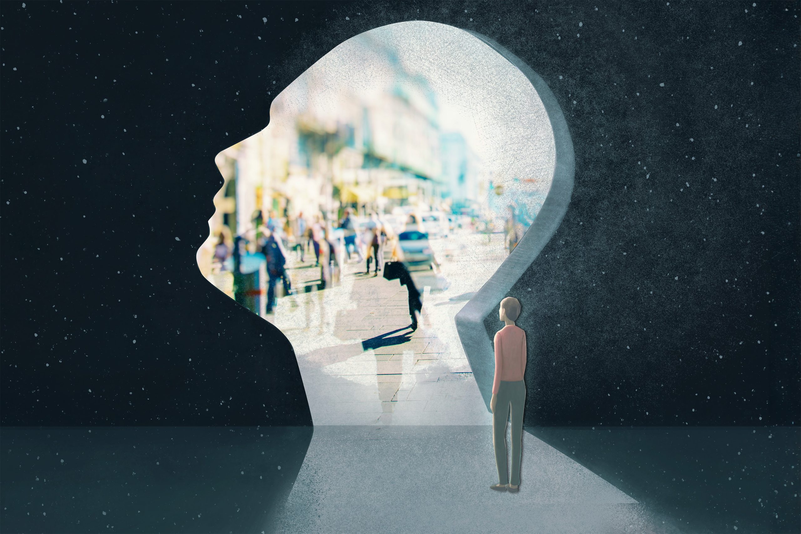
MIT neuroscientists have found that the brain uses the same cognitive representations whether navigating through space physically or mentally.

New research from MIT has found that such mental maps also are created and activated when you merely think about sequences of experiences, in the absence of any physical movement or sensory input. In an animal study, the researchers found that the entorhinal cortex harbors a cognitive map of what animals experience while they use a joystick to browse through a sequence of images. These cognitive maps are then activated when thinking about these sequences, even when the images are not visible.
This is the first study to show the cellular basis of mental simulation and imagination in a nonspatial domain through activation of a cognitive map in the entorhinal cortex.
“These cognitive maps are being recruited to perform mental navigation, without any sensory input or motor output. We are able to see a signature of this map presenting itself as the animal is going through these experiences mentally,” says Mehrdad Jazayeri, an associate professor of brain and cognitive sciences, a member of MIT’s McGovern Institute for Brain Research, and the senior author of the study.
McGovern Institute Research Scientist Sujaya Neupane is the lead author of the paper, which appears today in Nature. Ila Fiete, a professor of brain and cognitive sciences at MIT, a member of MIT’s McGovern Institute for Brain Research, and director of the K. Lisa Yang Integrative Computational Neuroscience Center, is also an author of the paper.
Mental maps
A great deal of work in animal models and humans has shown that representations of physical locations are stored in the hippocampus, a small seahorse-shaped structure, and the nearby entorhinal cortex. These representations are activated whenever an animal moves through a space that it has been in before, just before it traverses the space, or when it is asleep.
“Most prior studies have focused on how these areas reflect the structures and the details of the environment as an animal moves physically through space,” Jazayeri says. “When an animal moves in a room, its sensory experiences are nicely encoded by the activity of neurons in the hippocampus and entorhinal cortex.”
In the new study, Jazayeri and his colleagues wanted to explore whether these cognitive maps are also built and then used during purely mental run-throughs or imagining of movement through nonspatial domains.
To explore that possibility, the researchers trained animals to use a joystick to trace a path through a sequence of images (“landmarks”) spaced at regular temporal intervals. During the training, the animals were shown only a subset of pairs of images but not all the pairs. Once the animals had learned to navigate through the training pairs, the researchers tested if animals could handle the new pairs they had never seen before.
One possibility is that animals do not learn a cognitive map of the sequence, and instead solve the task using a memorization strategy. If so, they would be expected to struggle with the new pairs. Instead, if the animals were to rely on a cognitive map, they should be able to generalize their knowledge to the new pairs.
“The results were unequivocal,” Jazayeri says. “Animals were able to mentally navigate between the new pairs of images from the very first time they were tested. This finding provided strong behavioral evidence for the presence of a cognitive map. But how does the brain establish such a map?”
To address this question, the researchers recorded from single neurons in the entorhinal cortex as the animals performed this task. Neural responses had a striking feature: As the animals used the joystick to navigate between two landmarks, neurons featured distinctive bumps of activity associated with the mental representation of the intervening landmarks.
“The brain goes through these bumps of activity at the expected time when the intervening images would have passed by the animal’s eyes, which they never did,” Jazayeri says. “And the timing between these bumps, critically, was exactly the timing that the animal would have expected to reach each of those, which in this case was 0.65 seconds.”
The researchers also showed that the speed of the mental simulation was related to the animals’ performance on the task: When they were a little late or early in completing the task, their brain activity showed a corresponding change in timing. The researchers also found evidence that the mental representations in the entorhinal cortex don’t encode specific visual features of the images, but rather the ordinal arrangement of the landmarks.
A model of learning
To further explore how these cognitive maps may work, the researchers built a computational model to mimic the brain activity that they found and demonstrate how it could be generated. They used a type of model known as a continuous attractor model, which was originally developed to model how the entorhinal cortex tracks an animal’s position as it moves, based on sensory input.
The researchers customized the model by adding a component that was able to learn the activity patterns generated by sensory input. This model was then able to learn to use those patterns to reconstruct those experiences later, when there was no sensory input.
“The key element that we needed to add is that this system has the capacity to learn bidirectionally by communicating with sensory inputs. Through the associational learning that the model goes through, it will actually recreate those sensory experiences,” Jazayeri says.
The researchers now plan to investigate what happens in the brain if the landmarks are not evenly spaced, or if they’re arranged in a ring. They also hope to record brain activity in the hippocampus and entorhinal cortex as the animals first learn to perform the navigation task.
“Seeing the memory of the structure become crystallized in the mind, and how that leads to the neural activity that emerges, is a really valuable way of asking how learning happens,” Jazayeri says.
The research was funded by the Natural Sciences and Engineering Research Council of Canada, the Québec Research Funds, the National Institutes of Health, and the Paul and Lilah Newton Brain Science Award.


